Expert and Comfortable Oral Surgery for Impacted Teeth
If a tooth fails to erupt through the gums and remains deep in the bone, it’s considered impacted. The term impacted teeth is most associated with third molars (wisdom teeth); indeed, 10 million are extracted yearly in the U.S. due to impaction, infection, overcrowding, and pain. Wisdom tooth extraction in Spring Hill, Lecanto, Land O’ Lakes, and Clearwater, FL, is advised if you or a family member develops problems.
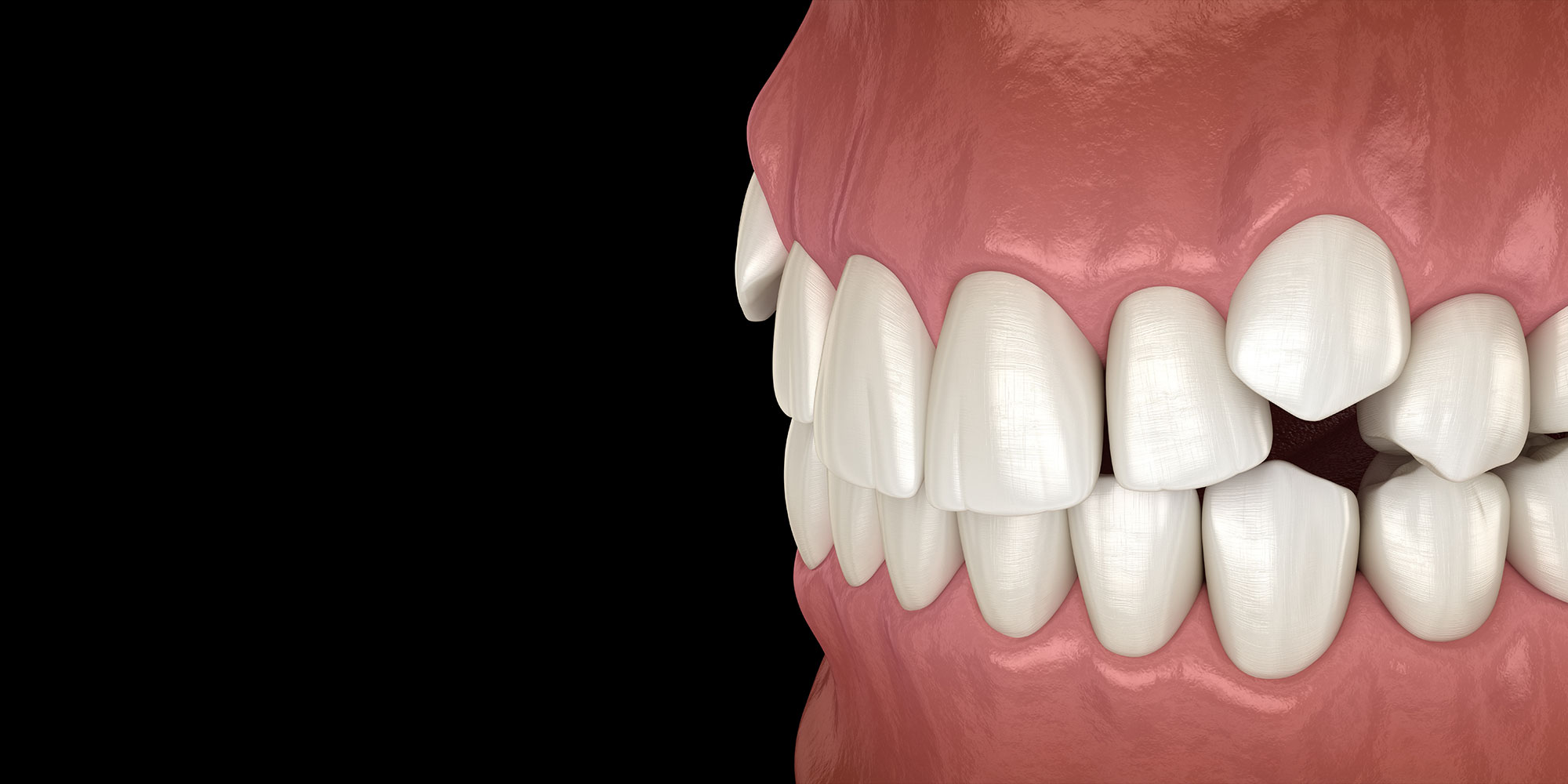
Maxillary canines (cuspids or upper eyeteeth) are the second most common teeth to become impacted. Unlike third molars at the back of your mouth, cuspid teeth are essential for chewing, speaking, and proper bite alignment, and they are clearly visible when you smile. Extracting a primary (baby) tooth may be advised to enable eruption of the permanent tooth while removing an impacted adult canine tooth and replacing it with a dental implant is a last resort when surgical procedures have failed.
Eruption and Anatomical Features of Maxillary Canine Teeth
Maxillary canines are the last of the “front” teeth to erupt into place normally. They usually come into place around age 13 and cause any space left between the upper front teeth to close together tighter. These teeth are extremely strong and designed to withstand the biting forces when your teeth come together. They also guide the rest of your teeth into a proper bite.
Incidence and Causes of Maxillary Cuspid Impaction
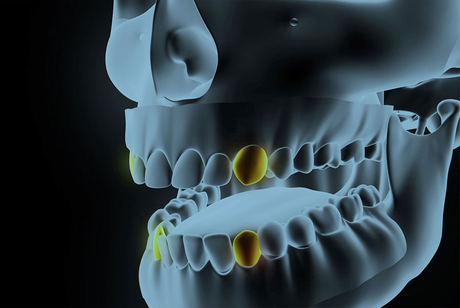
Maxillary cuspid impaction impacts an estimated 1% to 4% of the population. Sixty percent of these impacted teeth are located on the dental arch’s palatal (roof of the mouth) side. The remaining impacted teeth are found in the middle of the supporting bone—stuck in an elevated position above the roots of the adjacent teeth—or out to the facial side of the dental arch.
Studies suggest a combination of genetics causes maxillary canine impaction, comparatively longer tooth roots and path of eruption, development deep in the jaw, eruption after neighboring teeth, and lateral incisor abnormalities. Other documented causes of impacted maxillary canines include the presence of extra teeth (supernumerary teeth), odontomas (benign tumors), early trauma to the upper jaw, and cleft lip and palate.
Diagnosis and Treatment of Impacted Canines
The American Association of Orthodontists recommends that children have an orthodontic examination and panoramic radiograph (X-ray) by age 7 to monitor tooth eruption and detect any impactions. Dr. Michael Hashemian will perform a detailed assessment of the impacted tooth to pinpoint its location, angulation, and orientation.
A panoramic radiograph provides comprehensive information regarding the dentition, jaws, and surrounding structures with a lower radiation dose than cone-beam computed tomography. If a cuspid tooth gets impacted, we make every effort at Dentofacial & Cosmetic Surgery Institute to help it erupt into its proper position. Doing so can prevent issues such as painful and swollen gums, cysts, infections, and problems with adjacent teeth.
Does Age Matter?
Experts agree that an impacted maxillary canine will not erupt by natural forces alone in older patients, even if the space is available for the tooth to fit in the dental arch. Some studies analyzing surgical exposure/orthodontic treatment efficacy concluded that age matters, while others found no difference in success rates. We recommend early diagnosis and intervention to prevent dental problems, save time and money, and more complex treatment in permanent dentition.
Surgical Exposure of Impacted Canines
We offer general anesthesia and IV sedation in Spring Hill, Lecanto, Land O’ Lakes, and Clearwater, FL, to ensure comfort during extraction and surgery. If a primary (baby) tooth is present, this is extracted first. If any extra teeth, growths, or bone is blocking the path of eruption of adult teeth, these need to be removed.
Dr. Hashemian surgically exposes the tooth during the procedure to allow it to erupt. After the surgery, a protective dressing is placed over the surgical site while it heals. This method enables the canine tooth to emerge until it’s at the level of the adjacent teeth, after which you can get orthodontic treatment if needed.
Exposure and Attachment of Bracket and Chain
Dr. Hashemian frequently gets referrals from general dentists for many procedures and collaborates with orthodontists on surgical exposures. If your child or teen is under the care of an orthodontist and they request it, Dr. Hashemian will bond an orthodontic bracket to the exposed tooth. The bracket has a tiny gold chain temporarily attached to an orthodontic archwire.
In most cases, the gum tissue is placed back in its original location, and sutures are applied so only the chain remains visible as it exits a small hole in the gums. One to 14 days after surgery, the orthodontist attaches a rubber band to the chain. This exerts a light pulling force on the impacted tooth to help gradually move it into the proper position, which can take up to 12 months.